Pre-clinical and clinical facilities
HollandPTC has a variety of pre-clinical and clinical research facilities. There is an R&D proton beamline, a biology, and a physics lab. Furthermore, there are 2 treatment gantries and a dedicated treatment station for ocular tumours. These 3 treatment facilities used in clinical care can also be utilized for clinical studies or other R&D purposes. The imaging equipment consists of a CT scanner, a PET/CT scanner, and an MRI scanner. In addition, there is a research database with prospectively-collected patient information.
This database comprises health, treatment and follow-up information of all consenting patients treated at HollandPTC. Anonymized or coded data can be requested following the MREC-approved procedure for researchers at HollandPTC and its consortium partners.
Pre-clinical research facilities
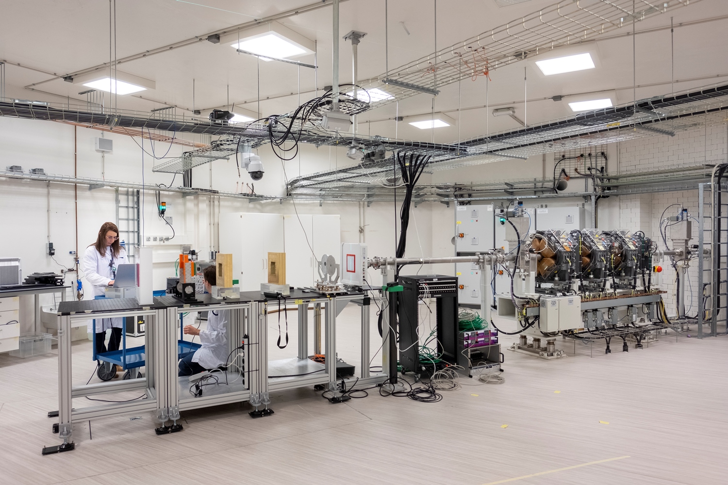
R&D proton beamline
The R&D bunker of HollandPTC is equipped with a fixed horizontal proton beamline, delivering a therapeutic proton beam with energies ranging from 70 MeV up to 250 MeV. The ProBeam superconductive cyclotron of Varian is able to deliver nominal beam currents from 1 nA up to 800 nA, with a transmission efficiency to the target position of 0.04% for the minimum energy up to 40% for the maximum energy. The therapeutic pencil beam with Gaussian profile in x and y is used for fundamental physics applications, device calibration and radiation hardness tests. Moreover, the beamline is equipped with a dual-ring passive scattering system which allows the production of large fields ranging from 2x2cm up to 20x20cm. Pristine Bragg peaks as well as Spread-Out Bragg peaks can be created. The passive system is well suited for radiobiological applications, such as in vitro and in vivo experiments.
Physics lab
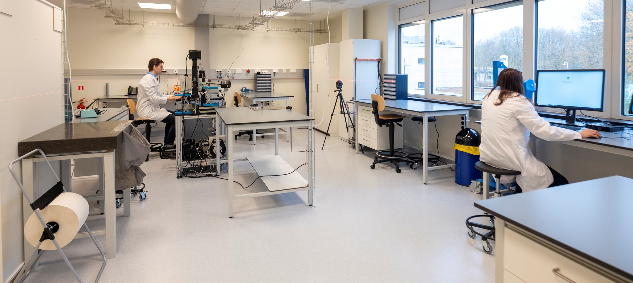
The physics laboratory is a classified D-Lab where radiological and chemical work can be performed. The lab offers the tools for preparing setups for irradiation in the R&D bunker, chemical work in a fume hood and the radiation safety equipment needed for independent radiological experiments.
Biology lab
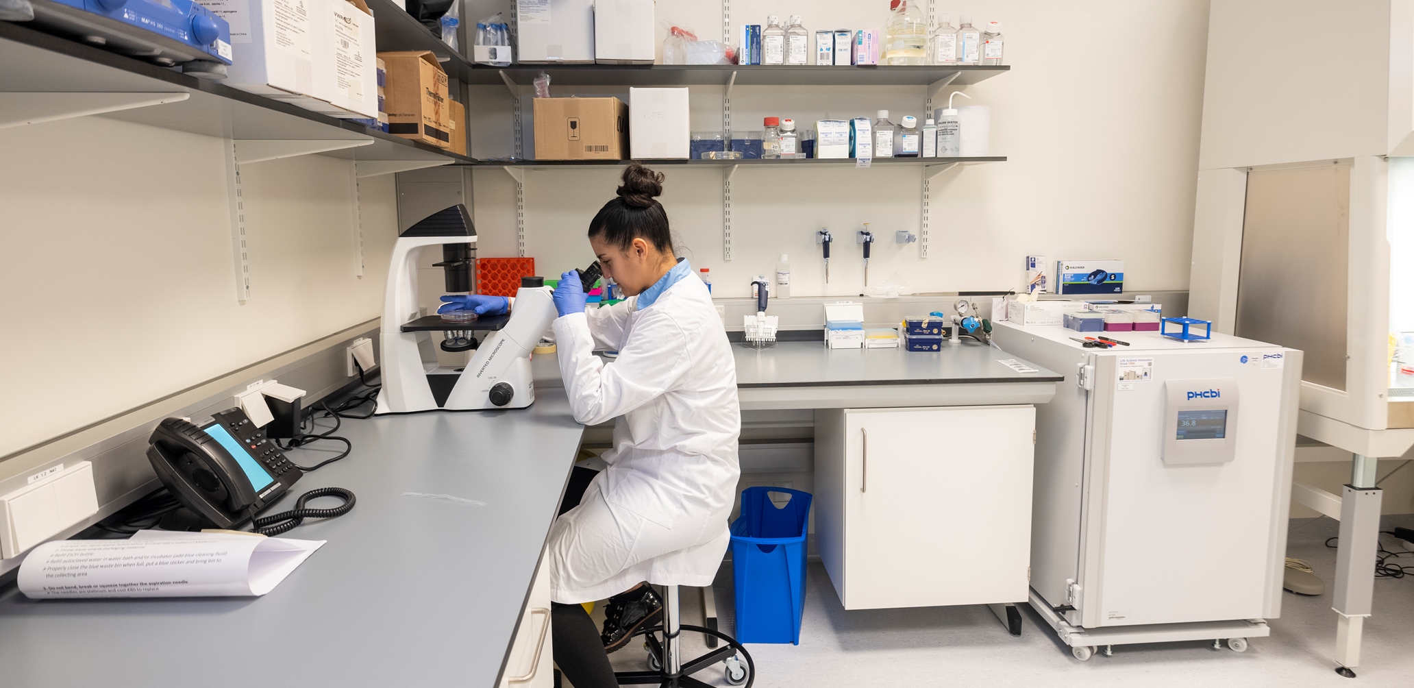
The biology laboratory is equipped with a confocal microscope, incubator, biosafety cabinet, freezer and fridge in order to perform basic cell culture work. Basic storage of consumables is also present. In the laboratory the biological specimen is prepared for proton irradiation and afterwards basic clonogenic survival analysis can be performed.
Clinical facilities
Treatment rooms
Treatment gantries
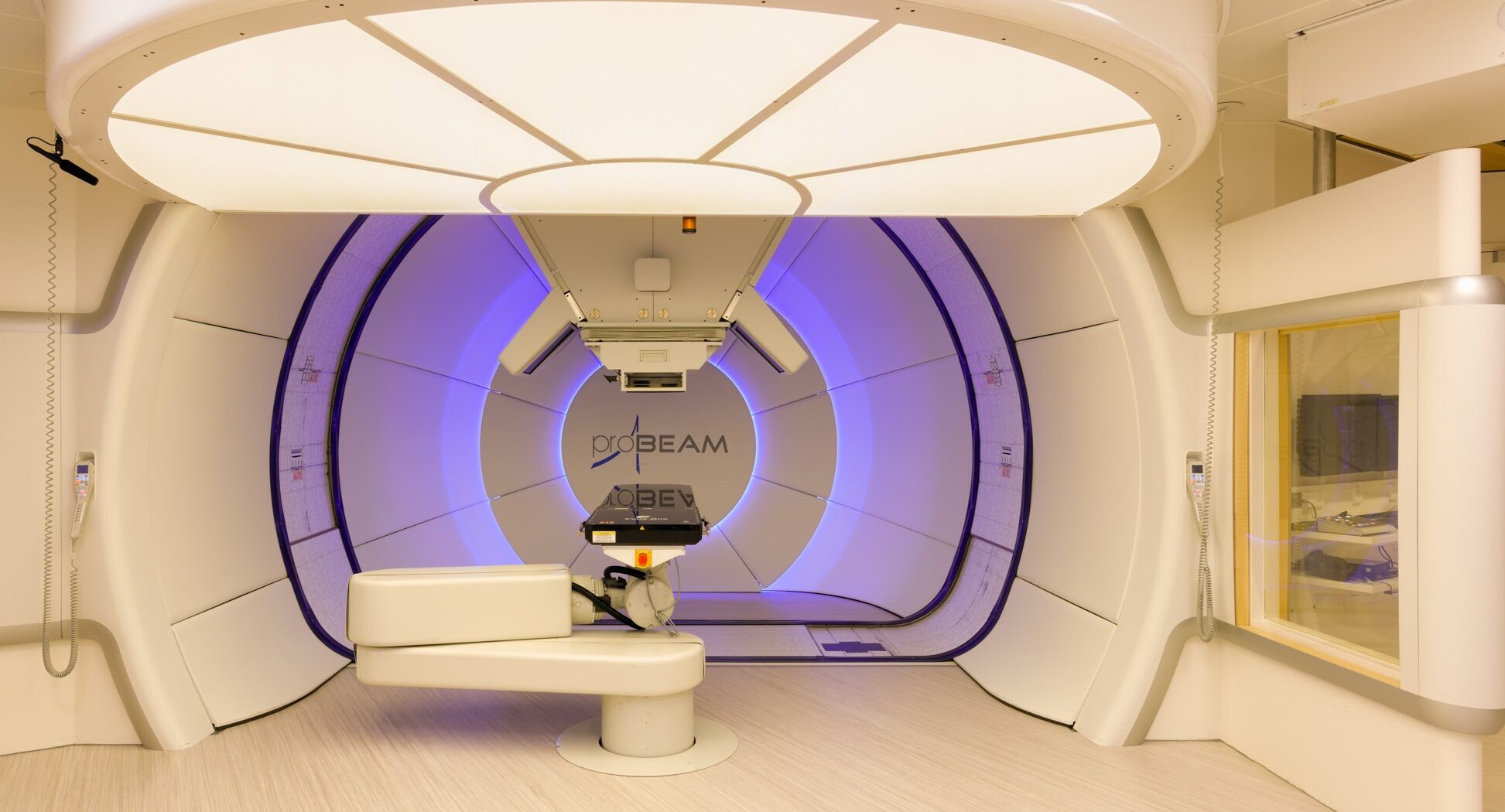
The 2 clinical proton therapy systems use pencil beam scanning technology in 360-degree gantries and are mounted with kV/cone-beam computer tomography (CBCT), for patient position verification. Furthermore, both treatment rooms are equipped with an in-room sliding Siemens SOMATOM Confidence CT scanner and a robotic bed to move the patient from imaging to treatment position, without altering the positioning.
Ocular station
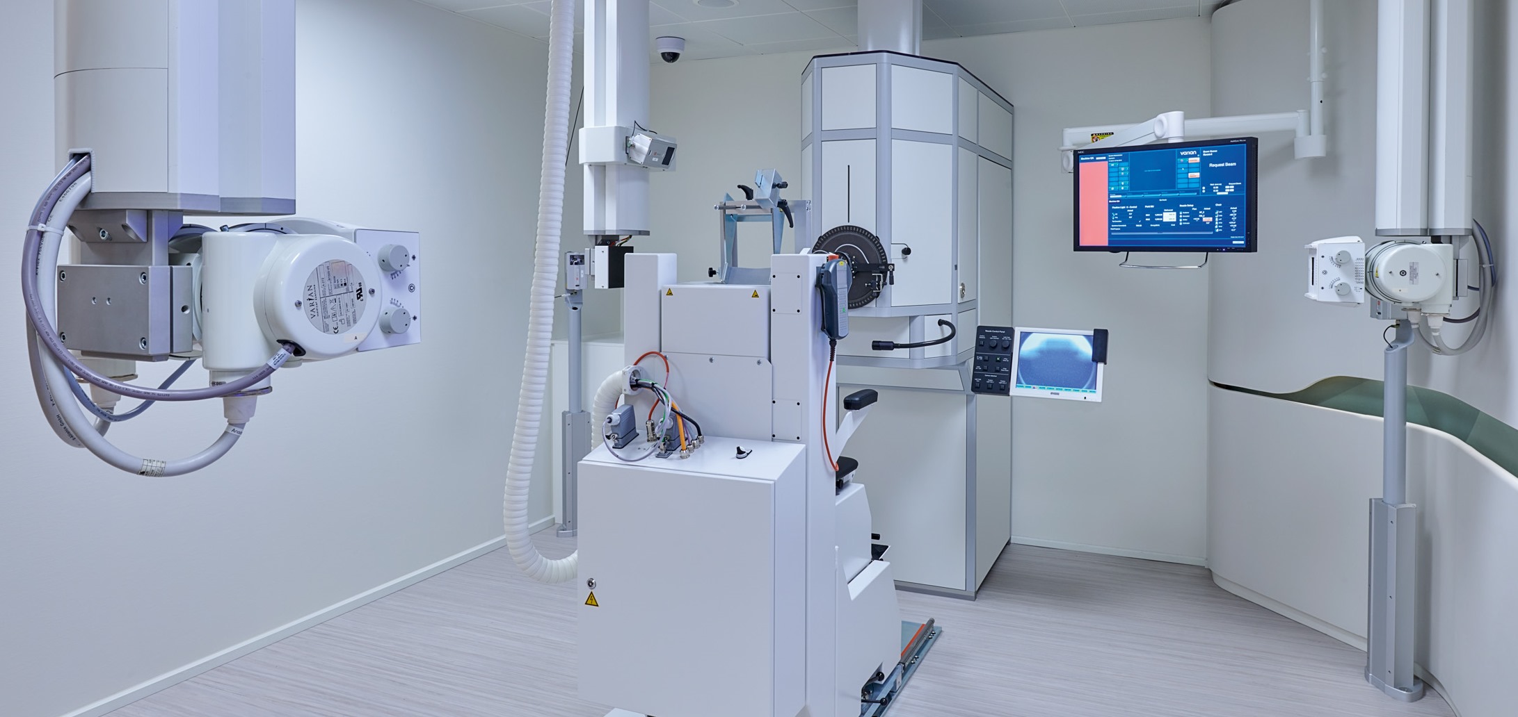
The ocular station consists of a fixed horizontal beamline and a single-scattering nozzle. It is fitted with orthogonal X-ray imaging equipment, utilizing digital flat-panel detectors, for patient position verification. A robotic chair allows positioning and immobilization of the patient with respect to the proton beam.
Imaging equipment
All imaging equipment is equipped with flat, hard-top couches and external lasers. Radiotherapy immobilization molds and indexing devices can be used.
CT scanner
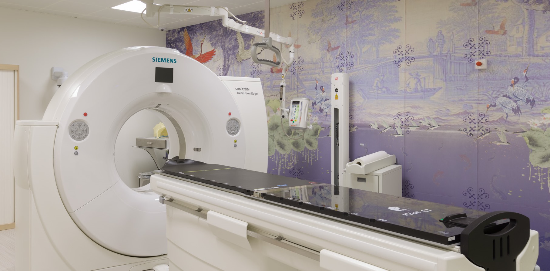
Siemens SOMATOM Definition Edge 128-slice dual-energy CT scanner, a single-source TwinBeam system with the option for simultaneous acquisition with 2 energy spectra for improved image quality and tissue characterization. This scanner is used for diagnostic imaging, treatment planning, treatment evaluation and treatment adaptation. It has 4D scanning options to detect motion and perform motion measurements.
PET/CT scanner
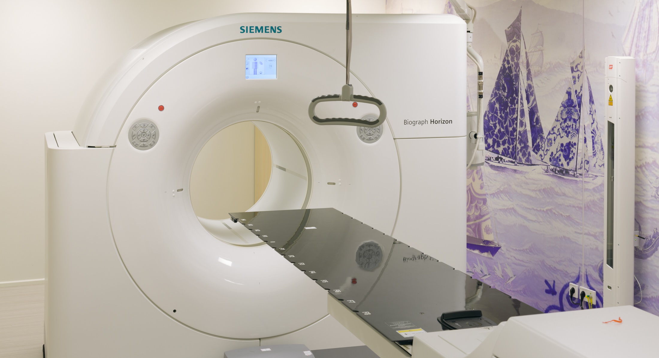
Siemens Biograph Horizon PET/CT scanner with TrueV axial gantry extension, with the option for imaging in treatment position including patient set-up based on external lasers. The scanner is used for diagnostic imaging, treatment planning and treatment follow-up.
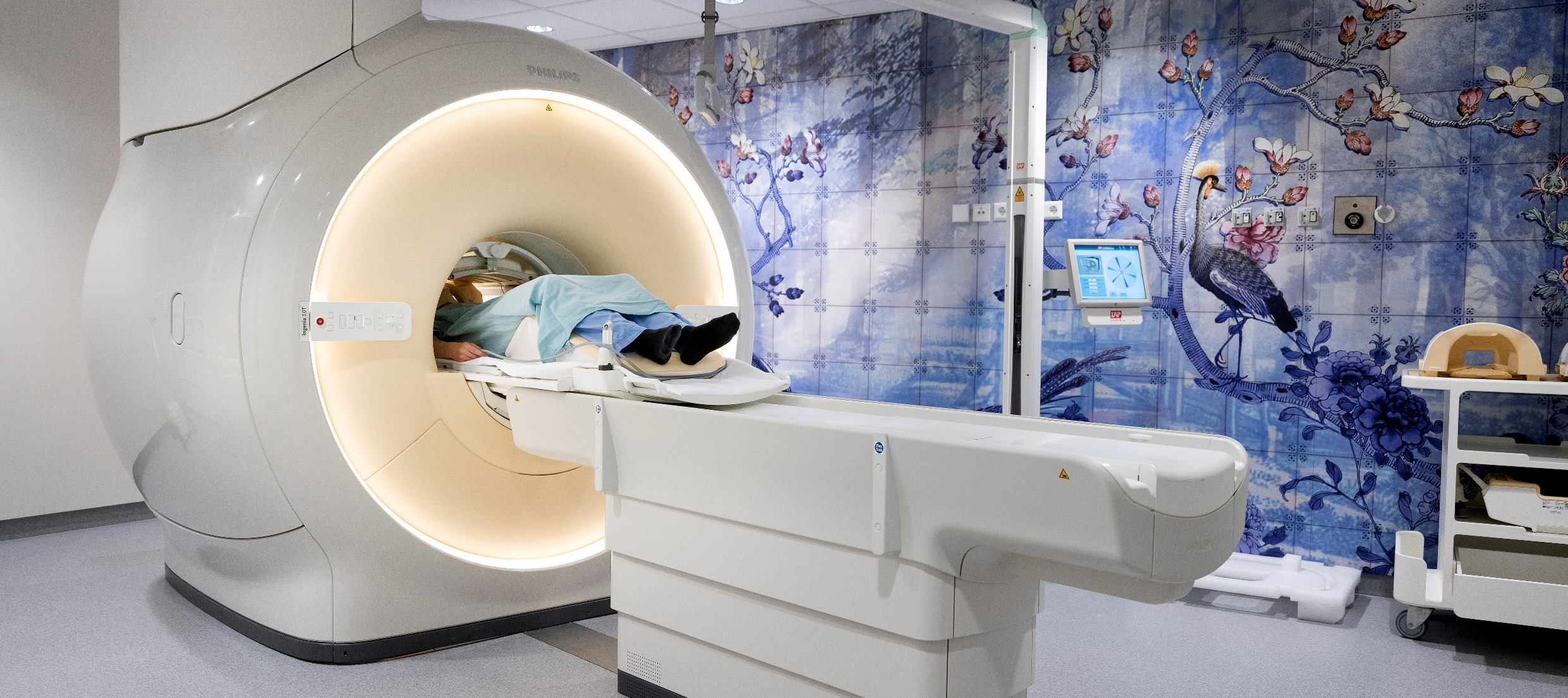
MRI scanner
Philips Ingenia MR-RT wide-bore 3T scanner dedicated for imaging in treatment position, including patient set-up based on external lasers. The scanner is used for diagnostic imaging, treatment planning and treatment follow-up.
